
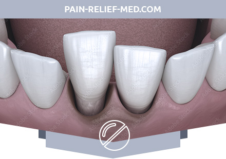
What is a Tooth Dislocation?
Dislocation of the tooth is damage to the ligamentous apparatus of the tooth, leading to displacement of the tooth in the hole, occurring in the lateral or vertical direction of the traumatic force. Dislocation often exposed frontal teeth of the upper jaw, less often the lower.
Causes of Tooth Dislocation
Dislocation of the tooth occurs under the influence of mechanical stress and is accompanied by damage to the ligamentous apparatus of the tooth.
Dislocations of the central teeth and canines often develop upon impact, fall; molars tend to dislocate with careless removal of the adjacent tooth.
When a tooth is dislocated, a fracture of the wall of the alveoli and the neurovascular bundle of the tooth can occur.
The cause of tooth dislocation is the force applied to its tooth crown:
– hit (injury);
– biting hard food;
– foreign body in chewed food;
– bad habits (opening bottles with teeth);
– inaccurate extraction of teeth, leading to dislocation near the standing tooth.
Pathogenesis during Tooth Dislocation
Dislocation of the tooth is the displacement of the tooth in the hole, which occurs when the lateral or vertical direction of the traumatic force. In a normal periodontal condition, considerable effort is required to displace the tooth. However, during bone resorption, dislocation can occur from hard foods and be accompanied by damage to the integrity of the gums. It can be isolated or in combination with a fracture of the root of the tooth, alveolar process or jaw body.
Symptoms of Tooth Dislocation
There are the following types of tooth dislocations:
– Complete dislocation of a tooth is characterized by dropping it out of the hole.
– Incomplete dislocation – partial displacement of the root of the alveoli, always accompanied by rupture of the periodontal fibers over a greater or lesser extent.
– Impacted dislocation is manifested by partial or complete displacement of the tooth from the hole in the direction of the jaw body, leading to significant destruction of bone tissue.
In case of incomplete dislocation of the tooth, the position of the tooth in the dentition changes. Complaints of pain, tooth mobility, changing its position in the dentition, impaired chewing function. When examining the oral cavity, incomplete dislocation of the tooth is characterized by changing the position (displacement) of the crown of the injured tooth in a different direction (orally, vestibular, distal, towards the occlusal plane, etc.). The tooth can be mobile and sharply painful with percussion, but not shifted beyond the limits of the dentition. The gums are swollen and hyperemic, it may rupture. Due to the rupture of the circular ligament of the tooth, periodontal tissues and damage to the wall of the alveoli, pathological dento-gingival pockets and bleeding from them can be determined. When a tooth is dislocated and its crown is displaced orally, the root of the tooth is usually displaced vestibularly, and vice versa. When the tooth is displaced in the direction of the occlusal plane, it protrudes above the level of the adjacent teeth, it is mobile and interferes with occlusion. Very often, the patient has a concomitant injury to the soft tissues of the lips (contusion, hemorrhage, wound).
Complications and outcomes of incomplete dislocation: shortening of the root of the tooth, obliteration or expansion of the root canal with the formation of intrapulpar granuloma, stopping the formation and growth of the root, curvature of the tooth root, changes in the periapical tissues in the form of chronic periodontitis, root cyst.
Complete dislocation of the tooth (traumatic extraction) occurs after complete rupture of the periodontal tissues and the circular ligament of the tooth as a result of a strong blow to the crown of the tooth. Most often affected frontal teeth in the upper jaw (mainly central incisors) and less often in the lower jaw.
The clinical picture of complete dislocation of the tooth: when examining the oral cavity, the tooth is absent in the tooth row and there is a bleeding of the dislocated tooth or filled with a fresh blood clot. Often there are related damage to the soft tissues of the lips (bruises, wounds of the mucous membrane, etc.). When referring to a dentist, sprained teeth are often brought in the pocket. To draw up a treatment plan, it is necessary to assess the condition of the dislocated tooth (the integrity of the crown and root, the presence of carious cavities, a temporary tooth or a permanent one, etc.).
Impacted dislocation (tooth intrusion) is the partial or total immersion of the tooth crown in the alveoli, and the tooth root in the spongy bone tissue of the jaw as a result of injury. As a rule, the front teeth on the upper jaw suffer with a strong impact on the cutting edge. When the dislocated impacted tooth is embedded in the thickness of the bone, periodontal fibers partially or completely ruptured, the cortical plate of the tooth hole is broken, especially in the bottom area.
Patients complain that after an injury, the tooth became shorter than the adjacent teeth or is not visible at all. A decrease in the height of the visible part of the tooth crown is clinically determined, the tooth is located above (below) the occlusal plane. The tooth is stable, its percussion is weakly painful. Sometimes the crown of the injured tooth is not visible at all, as it is completely immersed in the hole of the tooth. In this case, the examination of the hole reveals the cutting edge of the tooth. There is lune bleeding and gum mucous rupture.
Diagnosis of Tooth Dislocation
Diagnosis of tooth dislocation does not present any difficulties. Dislocation is determined by examination of the oral cavity on changes in the dentition, bleeding gums and abnormal mobility of the tooth.
Treatment of Tooth Dislocation
First of all, it is necessary to solve the problem of the advisability of maintaining such a tooth. The main criterion is the state of the bone tissue at the root of the tooth. If it is safe for at least 1/2 the length of the root, it is advisable to keep the tooth. First, the tooth is placed in its former place (under anesthesia), and then it is given rest, excluding its mobility. For this purpose, carry out splinting (wire or quick-hardening plastic). Then you should determine the condition of the tooth pulp. In some cases, when the root is displaced, the neurovascular bundle is ruptured, but sometimes the pulp remains viable. In the first case, with necrosis, the pulp must be removed, the channel must be sealed, in the second case the pulp is preserved. To determine the state of the pulp measure its response to an electric current. The reaction of the pulp to a current of 2-3 μA indicates its normal state. However, it should be remembered that in the first 3-5 days after the injury, a decrease in the excitability of the pulp may be a response to the traumatic impact. In such cases, it is necessary to check the state of the pulp in the dynamics (again). The restoration of excitability indicates the restoration of a normal state.
If the tooth responds to a current of 100 µA or more, it indicates necrosis of the pulp and the need to remove it. In the event of a tooth injury, it is possible to hammer the root into the jaw, which is always accompanied by rupture of the neurovascular bundle. This condition is accompanied by pain, and the patient points to a “shortened” tooth. In this case, the tooth is fixed in the correct position and the necrotic pulp is immediately removed. It is recommended to remove it as early as possible to prevent the decay and staining of the tooth crown in a dark color.
In case of acute injury, there may be a complete dislocation (a tooth is brought in the hands or a tooth dropped is inserted into the hole). Treatment consists of tooth replantation. This operation can be successful with unchanged periodontal tissues. Carry it out in the following sequence: trepan the tooth, remove the pulp and seal the canal. Then, after the root and the hole have been treated with antiseptic solutions, the tooth is introduced into place and fixed (in some cases splinting is optional). In the absence of complaints of pain spend monitoring and radiological control. The root of the tooth, replanted in the first 15-30 minutes after injury, is resorbed slightly, and the tooth is preserved for many years. If the replantation is performed at a later date, then the root resorption is radiologically determined within the first month after the replantation. Resorption of the root progresses, and by the end of the year a significant part of it is resorbed.
Treatment of incomplete dislocation of the tooth.
– tooth reposition;
– fixing with kappa or smooth splint;
– sparing diet;
– inspection after 1 month;
– when establishing the death of the pulp – extirpation of it and sealing the canal.
Immobilization or fixation of teeth is carried out in the following ways:
- Ligatural binding of teeth (simple ligature binding, continuous in the form of the eight, binding of teeth according to Baronov, Obwegezer, Frihoff, etc.). Ligature binding of teeth is shown, as a rule, in a constant bite in the presence of stable, adjacent teeth (2-3 in both directions from dislocated). For ligature tooth binding, thin (0.4 mm) soft bronze-aluminum or stainless steel wire is commonly used. The disadvantage of these methods of splinting is the impossibility of their use in the temporal bite for the above reasons. In addition, the imposition of wire ligatures rather time-consuming process. At the same time, this method does not allow sufficiently rigidly fix the sprained teeth.
- Tire-clip (wire or tape). The tire is made (bent) from stainless wire from 0.6 to 1.0 mm. thick or standard steel tape and fixed to the teeth (2-3 in both directions from the dislocated) with thin (0.4 mm) ligature wire. Tire-clip is shown in a constant bite, as a rule, in the presence of a sufficient number of stable adjacent teeth.
Disadvantages: trauma, laboriousness and limited use in the temporal bite. - Tire-kappa. It is made, as a rule, from plastic in one visit, directly in the patient’s mouth after reposition of the teeth. Disadvantages: bite separation and difficulty in carrying out EDI.
- Teething bushes. Shown in any bite in the absence of a sufficient number of supporting, including the adjacent teeth. They are made of plastic with reinforced wire, laboratory after removing the impression and casting the jaw model.
- The use of composite materials, with the help of which the wire arches or other splinting structures are fixed to the teeth.
Immobilization of sprained teeth is usually carried out within 1 month (4 weeks). At the same time, it is necessary to strictly observe the oral hygiene for the prevention of inflammatory processes and damage to the enamel of splinted teeth.
Treatment of complete tooth dislocation consists of the following steps.
– pulp extirpation and canal filling;
– replantation;
– fixing for 4 weeks with a kappa or smooth splint;
– mechanically sparing diet.
It is necessary to examine the tooth hole and assess its integrity. Radiographically, with complete dislocation of the tooth, is determined by the free (empty) hole of the tooth with clear contours. If the hole of the dislocated tooth is destroyed, then the borders of the alveoli are not determined radiologically.
Indications for tooth replantation depend on the patient’s age, his general condition, the condition of the tooth itself and its hole, on whether a temporary tooth or a permanent tooth root is formed or not.
Tooth replantation is the return of a tooth to its own well. There are single-stage and delayed replantation of the tooth. In case of simultaneous replantation in one visit, a tooth is prepared for replantation, the root canal is sealed and its own replantation is performed, followed by splinting. For delayed replantation, the dislocated tooth is washed, immersed in a saline solution with an antibiotic, and placed temporarily (before replantation) in a refrigerator. After several hours or days, the tooth is trepanned, filled and replanted.
The operation of the tooth replantation can be divided into the following stages:
- Preparation of the tooth for replantation.
- Preparation of the tooth hole for replantation.
- Actually replantation of the tooth and fixing it in the hole.
- Postoperative treatment and observation in the dynamics.
After 1-1.5 months after the operation of the tooth replantation, the following types of tooth engraftment are possible:
- Engraftment of the tooth by the type of primary tension through the periodontium (syndesmosis). This is the most favorable periodontal type of fusion, depending mainly on the preservation of the viability of periodontal tissues. With this type of fusion, the periodontal gap is determined on a control radiograph.
- Engraftment of the tooth by the type of primary tension through the periodontium (syndesmosis). This is the most favorable periodontal type of fusion, depending mainly on the preservation of the viability of periodontal tissues. With this type of fusion, the periodontal gap is determined on a control radiograph.
- Engraftment of the tooth according to the mixed (periodontal-fibrous-bone) type of fusion of the tooth root and the wall of the alveoli. On a control radiograph with such an adhesion, the line of the periodontal gap alternates with areas of its narrowing or absence.
In the long-term period (several years) after tooth replantation, resorption (resorption) of the root of the replanted tooth may occur.