
What is Omsk Hemorrhagic Fever?
Omsk hemorrhagic fever is an acute viral disease characterized by natural foci, fever, hemorrhagic syndrome and damage to the nervous system.
The first descriptions of Omsk hemorrhagic fever were made by local doctors in the Omsk region in the period from 1940 to 1945. (B.P. Pervushin, G.A. Sizemova and others). Since 1946, Omsk hemorrhagic fever has been isolated into an independent nosological form. It was found that the main reservoir of infection is a narrow cranial vole, and the carrier of the tick is D.pictus. Another route of transmission was contact. The disease occurred after contact with the muskrat (the local population knew about it, and even had the name “muskrat disease”). Natural foci of Omsk hemorrhagic fever were identified in the steppe and forest-steppe regions of the Omsk, Novosibirsk, Tyumen, Kurgan, and Orenburg regions. The reservoir of infection in nature is mainly water rat, red-backed vole, muskrat, as well as ticks of D.pictus, D.marginatus, which can transmit the virus to the offspring transovarially. Omsk hemorrhagic fever is non-contagious. No cases of human infection have been observed.
Causes of Omsk Hemorrhagic Fever
The causative agent of the Omsk hemorrhagic fever – a virus containing RNA, belongs to the family Togaviridae of the genus Flavivirus. It multiplies in the cytoplasm on the Golgi apparatus membranes and is found in the reservoirs of the endoplasmic reticulum; virus size does not exceed 40 nm, it has a spherical shape, cubic symmetry, is covered with a two-layer membrane, which contains lipids and antigenic proteins that determine the group-, type-and species-specific determinants. The virus is highly sensitive to treatment with liposolvents (chloroform, ethyl ether, sodium deoxycholate, detergents); it dies pretty quickly at room and higher temperatures, at 60 ° C dies in a few minutes and almost instantly when boiling. The virus is very well preserved in the frozen or dried protein substrate.
It is stored up to 7 months in 50% glycerol, and up to 2-4 years – in a lyophilized form. When treated with formalin (1: 500) at + 4 ° C for 2 weeks, the virus loses its infectivity, retaining its antigenic properties. Pathogenic for adult and newborn mice; 4–8 days after intracerebral infection, they develop meningoencephalitis. Serological variability of the virus was established, two serovariants – subtype I with Kubrin and Balatun strains, subtype II with Bogolyubovka and Guryev strains. In each of these groups, one strain was isolated from a person and one from Dermacentor marginatus ticks. The identified antigenic variants of the virus are associated with two types of ticks – D. pictus and D. marginatus.
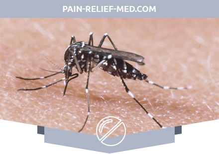
The main natural reservoirs of the virus in nature are the Ixodid mites Dermacentor pictus and Dermacentor marginatus. Established their spontaneous infection, the ability to transmit the virus along the course of metamorphosis, as well as transovarial their offspring.
Dermacentor pictus inhabits forest meadows, pastures, floodplain meadows, border steppe areas, it is a three-mite mite. Adult large mites parasitize on sheep, cows; young – on small mammals, often on voles; all forms – on hedgehogs and hares. He rarely attacks a person. Adult ticks are active in spring (in May) and in autumn (5% of spring activity), young in summer. In the Transcaucasus, their parasitism continues until winter.
Characteristic of Dermaсentor ticks is their different numbers on different pastures. The diversity of distribution of mites on pastures, and not on the territory in general, depends on the placement of voles on them – the owners of the young forms of mites. The springtime prevalence of sexually mature ticks on any plot corresponds to the summer distribution of the common vole population in previous years. The main biotopes are bumpy meadows, cuttings, forest edges, that is, places where voles can hide from predators. In addition, ticks are usually more where the cattle stay longer (“afternoon snack” places). Less ticks are observed where vegetation has been trampled (places where cattle are often driven). The indicated dependence of the number of ticks on the number of rodents can be used to predict the number of ticks. After the peak of the number of rodents on pastures, we can expect the mass appearance of ticks. Huge stocks of mature ticks that occur in nature in the year of mass reproduction of mouse-like rodents remain for the next 3-4 years.
Dermaсentor marginatus is found in the south of the European part of Russia, Transcaucasia, Kazakhstan, in the mountains of Central Asia, in the south of Western Siberia, in plains and mountain steppes, forest-steppe, mountain forests (pastures), beams, in floodplain meadows, in forest belts – in places where the possible owners live. Characteristically, D. marginatus is absent in populated areas. Adults feed on large domestic and wild mammals (wolves, hares, hedgehogs), they can attack humans; young forms parasitize small mammals – rodents, insectivores. Three-host species, parasitizing season – spring (from February to May in the south), partly autumn, for young forms – summer.
In nature, muskrats, water voles, housekeeper voles, narrow skull voles, shrews also serve as reservoirs of the pathogen in nature. Participation in the circulation of the virus and some other mammals (gophers, hares, hedgehogs, hamsters), birds (crows, rooks, bitterns), frogs and lizards is not excluded. In these animals, spontaneous infection and susceptibility to the virus were established under the experimental conditions. In animals there is both an acute form of the disease, with a fatal outcome, and latent infection.
The virus is more consistently excreted in the urine and feces of animals, which leads to infection of the environment. It is possible for a human to be infected by the alimentary route through water and food products infected with the secretions of sick rodents. In this case, the disease can occur in winter.
Spontaneous virus infection has been established in some species of mosquitoes (Mansonia richiardii, Aedes excrucians, Aedes flavescens, Culex modestus). Under certain conditions, they can participate in the transmission of the virus to animals as well as to humans.
Along with the transmissible and alimentary route, animals and humans can become airborne by inhalation of vaccinated material that enters the air when urine and feces of sick animals dry out.
In natural water bodies in the summer, the Omsk hemorrhagic fever virus is able to remain viable for 18–20 hours in concentrations sufficient for alimentary infection of small mammals.
Pathogenesis during the Omsk Hemorrhagic Fever
The gate of infection is the skin at the site of a tick bite or minor skin lesions infected by contact with muskrat or water rat. At the site of the gate infection of primary affect is not observed. The virus enters the bloodstream, hematogenously spreads throughout the body and mainly affects the blood vessels, nervous system and adrenal glands. At necropsy of those who died from Omsk hemorrhagic fever, a sharp plethora and swelling of the brain and spinal cord, serous hemorrhagic leptomeningitis, small hemorrhages, necrosis and focal encephalitis are revealed, sympathetic ganglia of the neck are also affected, the solar plexus, focal encephalitis, the sympathetic ganglia of the neck, the solar plexus, intervertebral patterns, and intervertebral patterns are affected. Pathological changes are similar to those of other hemorrhagic fevers.
Susceptibility to the Omsk hemorrhagic fever virus is universal and high. In the blood of patients with 2–3 days, complementary binding is formed, and from 14–20 days neutralizing antibodies persist for 1 to 3 years.
Most often, people who were in the field or forest (agricultural workers, hunters involved in the muskrat) are ill. The disease is sporadic or group in nature. Antibodies are often found in the blood of local residents; therefore, visitors often fall ill. After an illness, persistent immunity develops.
Symptoms of Omsk Hemorrhagic Fever
The incubation period often lasts from 2 to 4 days. Prodromal phenomena are rarely observed. The disease begins suddenly, the body temperature rises and as early as the first day reaches 39-40 ° C. There are general weakness, intense headache, pain in the muscles of the whole body. Patients are inhibited, reluctant to answer questions, lie on their side with their heads thrown back. Body temperature is kept at a high level for 3-4 days, then slowly lytically decreases by the 7-10th day of illness. Fever rarely lasts less than 7 or more than 10 days. Almost half of the patients have recurrent fever waves (relapses), more often at 2-3 weeks from the onset of the disease and last from 4 to 14 days. The total duration of the disease from 15 to 40 days.
When viewed from the 1-2 day of illness, almost all patients develop a hemorrhagic rash. The skin of the face, neck and upper parts of the breast is hyperemic, the face is puffy, the vessels of the sclera are injected. Appear nosebleeds, bleeding from the nasopharynx, pulmonary, intestinal, uterine. Subcleral hemorrhages are frequent. Hemorrhages are noticeable on the mucous membrane of the pharynx, on the gums. The skin has a profuse hemorrhagic rash from petechiae to major hemorrhages, hemorrhages in the sacrum can turn into extensive areas of necrosis. There is a decrease in blood pressure, deafness tones, possible bradycardia, pulse dicrotism and some extrasystoles. Approximately 30% of patients develop pneumonia (small focal), there may be signs of kidney damage. On the part of the central nervous system, signs of meningitis and meningoencephalitis (with severe forms of the disease) are noted. In the blood – severe leukopenia (1200-2000 in 1 μl), ESR is not increased.
Diagnosis of Omsk Hemorrhagic Fever
The diagnosis takes into account the epidemiological background (stay in an endemic area, seasonality, tick attack, contact with rodents, morbidity, etc.) and characteristic clinical manifestations (sudden onset, early manifestation of hemorrhagic syndrome, etc.).
To confirm the diagnosis, CSC, neutralization and passive hemagglutination reactions are used. In the early days of the disease, a virus can be isolated from the blood. For serological studies using paired sera of the patient, taken with an interval of 10-15 days. The virus is highly pathogenic for newborn and adult white mice with intracerebral and any other infection; the highest titers are found in the brain of newborn mice. The virus can also be passaged in cerebral transplants in young white rats. Experimental infection of the muskrat through the passage (from mice) of the Omsk hemorrhagic fever virus causes a highly contagious acute illness with hemorrhagic enteritis, pneumonia and frequent death. The process is less acute in water rats and young narrow-craned voles. Paralytic disease after intracerebral infection develops in monkeys of several species, suckers of white rats, young narrow cranial voles and in about 10% of cases in young guinea pigs. Many other vertebrates respond to experimental infection with the Omsk hemorrhagic fever virus only by short-term viremia and the development of specific antibodies without visible signs of disease.
The Omsk hemorrhagic fever virus multiplies relatively well in several types of tissue cell cultures (embryos of chickens, hamsters, mice, monkeys, humans), but only in cell cultures of pig embryo tissue gives a pronounced cytopathogenic affect with the destruction of a cell monolayer. It forms plaques – negative colonies of the virus under the agar layer in the cell cultures of embryos of pigs, hamsters and chickens. The Omsk hemorrhagic fever virus is identified in the RSK, in experiments neutralizing immune and hyperimmune homologous sera, in the reactions of suppressing hemagglutination and diffusion precipitation in agar. In all these reactions, there is a high level of group antibodies, common to all members of the tick-borne encephalitis antigen complex, but with a marked predominance of vortices in homologous systems. Only by pre-depleting group antibodies in immune sera (using dosed adsorption by a heterologous virus) can we obtain strictly specific results for identifying an infection caused by the Omsk hemorrhagic fever virus.
Using the fluorescence reaction, it is possible to detect and simultaneously identify the antigen of the Omsk hemorrhagic fever virus in infected tissue culture cells. In addition, the culture fluid of infected tissue cultures can be used to detect and identify the hemagglutinins of the Omsk hemorrhagic fever virus in the DSA and PHA. A study of early and late serum samples of a patient in the RSK, RPGA, RDPA or PH may confirm the diagnosis “Omsk hemorrhagic fever” if the second sample indicates the presence of antibodies to the Omsk hemorrhagic fever virus or 4-fold (or more) increase in their titers. Serological reactions such as RPHA and RDPA, which allow mass screening of people and animals for antibodies to the Omsk hemorrhagic fever virus, are successfully used to study serological epidemiology in areas endemic to Omsk hemorrhagic fever. Differentiate from other hemorrhagic fevers, tick-borne encephalitis.
Treatment of Omsk Hemorrhagic Fever
Treatment of Omsk hemorrhagic fever is exclusively symptomatic, with obligatory observance of strict bed rest for the patient. In case of bleeding, measures are taken to replace lost blood. Recommended hemostatic drugs, toning vascular wall, according to indications – antishock and heart. In addition, appropriate antibiotics are administered for inflammatory complications (in the throat and internal organs), which may develop a second time due to local hemorrhages. With the development of thrombohemorrhagic syndrome, intravenous administration of heparin 10 000-40 000 IU per day is used.
Prevention of Omsk Hemorrhagic Fever
Preventive and anti-epidemic measures are to carry out disinfestation, kill ticks and protect people from their attack, vaccination with killed formol vaccine. Due to the antigenic proximity of the virus of Omsk hemorrhagic fever and tick-borne encephalitis, the vaccine causes the formation of strong immunity against both diseases. Preventive measures should include primarily the extermination of water rats and other small animals on muskrats. In order to break the ecological contacts and interrupt the epizootic chain that circulates the pathogen in the biocenosis, it is necessary to systematically suppress the migration activity of small animals. In the conditions of lake basins, the use of plow furrows and long-lasting fighter points (ECD) are effective.
The plow furrow method is based on the use of high mobility of water rats and other small animals and is intended for their extermination. Furrow with the help of mounted tractor plow, specially converted for this purpose, is laid around the reservoir and supplied with trap cylinders. When moving animals use the furrow as a track, run along it and get into the cylinders. Thus, mammals of the dwelling and lake-marsh complex are caught for several months (from April to November), which significantly reduces the number of animals and prevents the transportation of the virus to the muskrat population.
ATCM are intended for the extermination of water rats and small animals in muskrat grounds. These are square boxes measuring 13x13x8 cm with semicircular inlets about 5 cm in size through which only water rats and smaller animals penetrate. Muskrats, ferrets, columns, ducks and other game animals do not pass into them. Such houses, pollinated inside 10 g of zinc phosphide, are installed on muskrats and in alloys. Pollinating ITD with zinc phosphide is recommended no more than once a year. Recycling ITDD this drug is advisable to carry out in May and June, that is, at the beginning of the increase in migratory activity of animals. With the help of ITDD, water rats and vole housekeepers are exterminated in their optimal habitat (on splavins) and muskrats. Using the plow furrow and ITDD method, the occurrence of epizootic among muskrats in small reservoirs (up to 1000 ha) can be prevented.